Mobile EEG combined with human eye and motion tracking – Gamze Adaçay, Turkey
Methods for measuring brain activity were clearly presented, followed by examples of experimental setups and studies. In the main part of the course we examined the question of why and how EEG became one of the most successful approaches in human neuroscience. To improve understanding, the subject was presented with various studies and images, including videos. The session was fluent, attractive and informative.
Super-resolution iPSC-derived sensory neurons – Anamaria-Domnica Vladoiu, Romania
In the iPSC (induced pluripotent stem cells) method class, we were guided through the principle and protocol of expansion microscopy, with the aim of studying iPSC-derived sensory neurons (iSN). We visualized cell cultures to check neuronal differentiation, went through phases such as gelation and chamber coating, and ultimately the high-resolution microscopy was impressive.
Basic introduction to genomic databases and computer-assisted sequence analysis – Tierney Kuhn, USA
The lecturer packed an incredible amount of information into just four hours. And crazy enough, I was able to follow! He started by showing us how to zoom into sequences of interest in UCSC's genome browser, to predicting their protein translation, to guiding us on how to design our own plasmids. Despite this fast pace, I never felt like I was lost because at every step he personally checked on each of us to make sure we were following him. Even when my computer crashed and I had to borrow my roommate's Chromebook, he found a way to get ApE running on my computer.
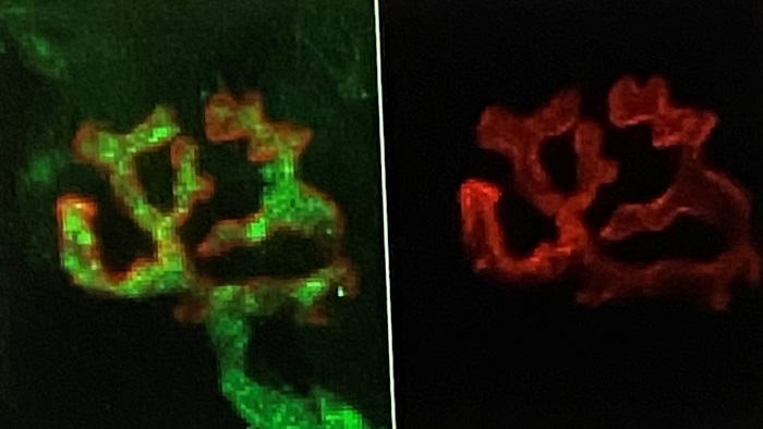
Immunocytochemistry of the neuromuscular junction – Camille Nicole Cauchi, Malta
After an informative overview of the experiments to be carried out, the tissue necessary for observing the neuromuscular connection was prepared. The neuromuscular junctions were stained and analyzed under the confocal microscope. The resulting images showed the expression of ThY1-YFP in selected neurons and provided a comprehensive view of the postsynaptic environment, as shown in the figure below.
During this session, we became familiar with novel methods and received detailed instructions on how to conduct the experiment. The integration of theoretical knowledge with practical application improved the understanding of the topic and demonstrated that this session is a great component to the overall learning experience.
A complete report on other course parts can be found on the homepage of the Elite Graduate Program “Translational Neuroscience”.
Text: Dr. Manuel Nagel, Gamze Adaçay, Anamaria-Domnica Vladoiu, Tierney Kuhn, Camille Nicole Cauchi


